We have introduced a new whitening system WHITE smile Flash
It is the latest LED technology, which has a step-by-step…

Today, cone beam computer tomography is an integral diagnostic service of a modern dental clinic.
 “Dubnova’s Clinic “STOMATOLOGiYA” always goes in the vanguard of dental technologies, methods and equipment, offering its clients diagnostic testing on a cone beam computer tomograph. This provides an opportunity to see the notable difference and create a comfort for the clients of the clinic.
Cone beam computer tomography or more simply CBCT of teeth is performed when you need to see the anatomical features of the structure of the teeth (for example, diagnose the presence / absence and position of the third that have not yet erupted), teeth primordia, dental roots, canals and state of bone tissue of the jaws, as well as to confirm a preliminary diagnosis.
CBCT is also used to estimate a level of the bone tissue while treating the gums.
If the teeth are in the wrong position, CBCT is performed before the start of the orthodontic treatment in order to visualize the location of the roots and the impacted teeth.
In case of trauma of the maxillofacial area, CBCT shows fractures of the bones, jaw displacement, damage to the roots of the teeth, and other problems that cannot be detected by visual inspection.
To treat the canals of the tooth, CBCT can: find additional canals, diagnose inflammatory processes, and estimate the quality of previous treatment and the features of anatomy before starting treatment.
In the case of neoplasms in the oral cavity, the CBCT comes to help the doctor to assess the size and prevalence of the inflammation area.
When planning dental surgery, CBCT can assess the anatomical features of a specific zone and predict the duration and extent of the operation.
The results of dental cone beam computer tomography are used in therapeutic, surgical, orthopedic, orthodontic treatment and in pediatric dentistry.
Cone beam computer tomography (CBCT) of teeth is the stage of complex diagnosis of teeth. Having precise data, the team of specialists determines all existing problems and selects the most effective treatment. However, cone beam computer tomography data can be useful not only for dentists. For example, a snapshot of the maxillary sinuses can be useful for check-up with an otolaryngologist (ENT).

A cone beam computer tomography is performed by applying the same principle that the muscles, soft tissues and bones of our body differently pass X-rays. The X-ray passes through the body and is captured by a special detector. The computer records the signals coming from the detector, converting them into a series of pictures, on the basis of which the computer builds a 3D model – a CBCT scan.
The scanning procedure itself is absolutely painless and lasts 0.7 – 19 seconds. (depending upon the type of examination) and does not require any preparation.
The result of the examination in digitized form can be recorded on any data medium (disk, USB flash drive) or downloaded to a file hosting service.
VATECH PaX-i3D Smart – a model of the latest generation of TECH & E-WOO, South Korea – the world leader in the production of tomographs, is used in our clinic.
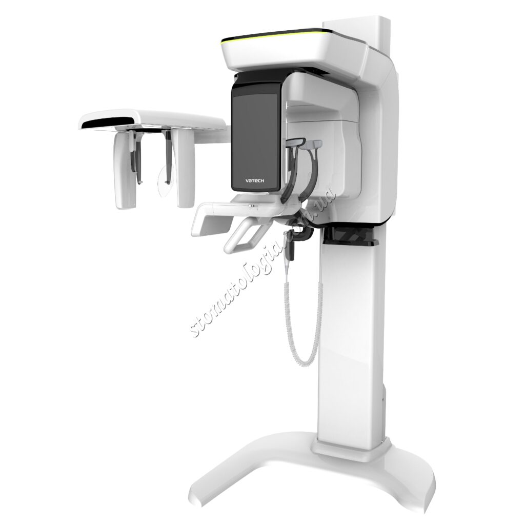
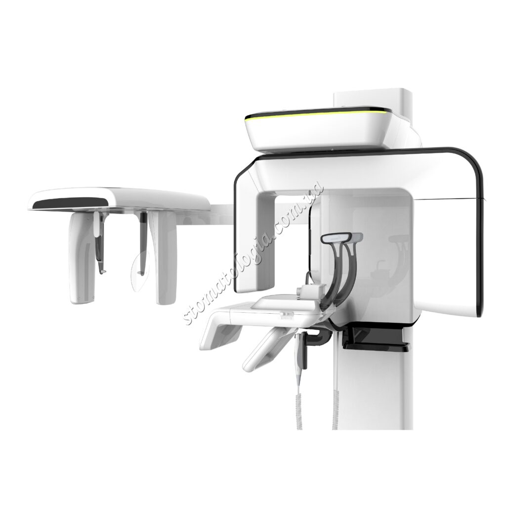
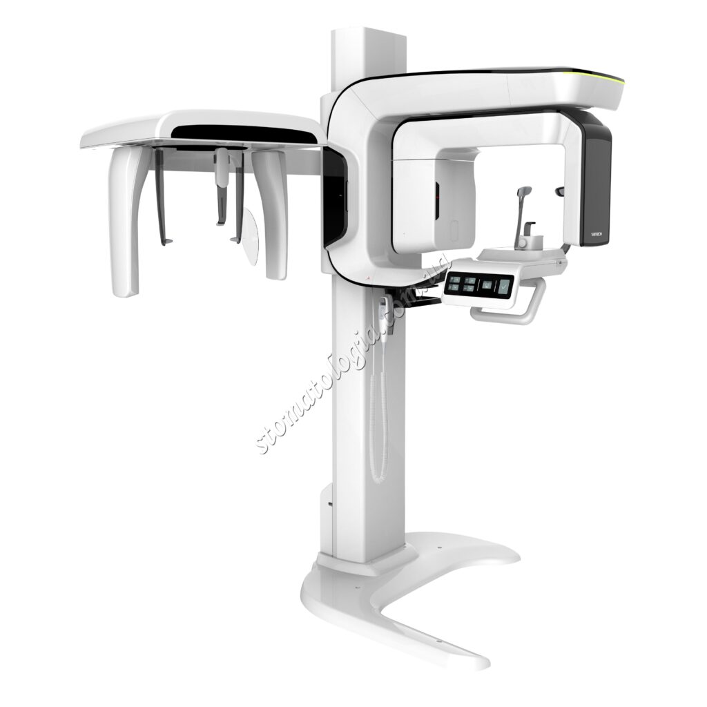
Photo 1. Computerized dental tomograph VATECH PaX-i3D Smart
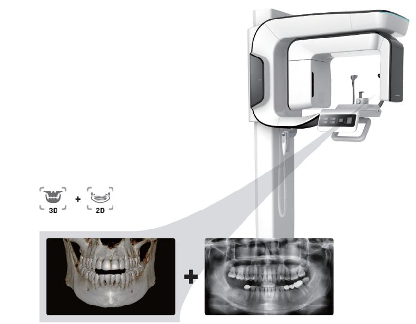
Photo 2. One scan – 2 images

Photo 3. The dose of X-ray exposure at the VATECH PaX-i3D Smart tomograph and standard (spiral) tomography

Photo 4. 3D visualization and simulation for implantation of teeth
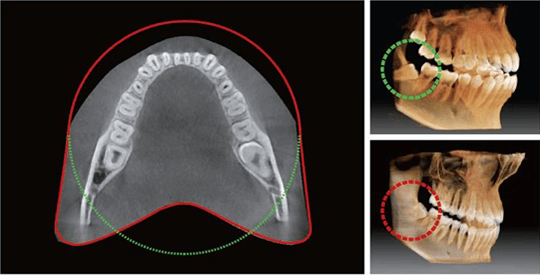
Photo 5. Wide view angle of the dentition, no hidden zones.
Cone beam computer tomography of the jaws is the most objective and reliable method of diagnosis in modern dentistry.

It is the latest LED technology, which has a step-by-step…

24 серпня остаточно перетворилося в найголовніше свято України. Сьогодні ми…

Минуло 500 днів широкомасштабної війни, і кожен з нас пережив…

365 days of struggle, pain, tears, hope and faith. The…

From all the employees of the clinics, we congratulate our…

Denis Viktorovych Podilchuk spoke at a dental conference with the…
2 jaw
Panoramic
Wide viewing angle of the dentition, no hidden zones.
3D visualization and simulation for dental implantation